Home
/ Amoeba Under Microscope 400X, Bio385 Protozoa, Images of amoeba cell under microscope www industrious info.
Amoeba Under Microscope 400X, Bio385 Protozoa, Images of amoeba cell under microscope www industrious info.
Amoeba Under Microscope 400X, Bio385 Protozoa, Images of amoeba cell under microscope www industrious info.. Biology protista amoeba malaria paramecium spirogyra. Although students can also see amoeba in the sample, it is possible to differentiate them from euglena. The word pseduopod means false foot. the pseudopod has two functions, or uses: Education and information about the brain eating ameba naegleria fowleri that causes encephalitis and death including frequently asked questions, biology, sources of infection, diagnosis, treatment, prevention and control, and other publications and pertinent information for the public and medical professionals. Image courtesy pearson scott foresman.
Paramecium captured under the microscope at 400x. Amoebas are simply single celled organisms. Bakare works for the fresh water protozoan diversity around wai and captured this vorticella video under the microscope by using a 2 megapixel eyepiece camera at 45x magnification. Moss captured under a biological microscope at 400x. It is just as if you put a ruler under.
Amoeba Protozoan Videos Olympus Ls from static4.olympus-lifescience.com Proteus may appear in a variety of colors (often yellow, green and purple) under a microscope. Here, students will observe them (euglena) as elongated organisms with a whip like tail on one end. It is just as if you put a ruler under. You may also want to learn more about actinophrys (small, heliozoan protists) here. When i place water samples on the microscope slides i usually wait a few minutes for the amoeba to attach to the glass and start moving. Arcella vulgaris (shown in the video above) is a testate amoeba. Under darkfield illumination at a magnification of 400x with a playing time of 37.3 seconds. First the colorless ectoplasma moves in front of the pseodopodia followed by the grained entopl.
Moss captured under a biological microscope at 400x.
And, in a culture with 50 or more amoeba proteus species, they can be easily seen in one field of view on a low power stereoscope as well. Field of view or field diameter is very important in microscopy as it is a more meaningful number than magnification. Although students can also see amoeba in the sample, it is possible to differentiate them from euglena. Amoebas are simply single celled organisms. Amoeba using its pseodopodia to ooze forward (100x magnification, no sound). Movement of the amoeba is shown. Secondly, what magnification do you need to see amoeba? The protozoa you will observe today is called the amoeba. Field diameter is simply the number of millimeters or micrometers you will see in your whole field of view when looking into the eyepiece lens. Paramecium under microscope 400x labeled. An amoeba's cell's organelles and cytoplasm are enclosed by the membrane. A large and diverse group of protozoa, flagellates are grouped by how they move. Hanny s voorwerp official website of discoverer hanny van arkel.
Pond water contains a number of arthropods such as copepods water fleas and ostracods crustaceans. Once called slipper animalcules due to their oblong shape, paramecium live in a variety of watery environments, both fresh and salt, although they are most abundant in stagnant bodies of water. Plantar warts are miniature growths appearing most commonly on the heel or other areas of feet or hands. New compound microscope donated to the lab soundbio lab. You may also want to learn more about actinophrys (small, heliozoan protists) here.
Lab Manual Exercise 1 from www2.palomar.edu Video recorded by lee beavington at. The protozoa you will observe today is called the amoeba. Hd amoeba at 40x 100x 200x and 400x youtube. Paramecium under microscope 400x labeled. If you are using a protozoa culture kit, protists will usually begin to appear after 24 hours with the most variety after about 3 days. Amoeba 400x as viewed by differential interference microscopy (dic) microsopy. Amoeba are shapeless (they look like a big blob) unicellular organisms from the genus protozoa. Believe it or not, this is a single cell!
Arcella under the microscope at 400x.
Under darkfield illumination at a magnification of 400x with a playing time of 36.4 seconds. Amoeba (plural amoebas/amoebae) is a genus that belongs to kingdom protozoa. Luke milbocker captured these images of protozoans using a compound microscope. A large and diverse group of protozoa, flagellates are grouped by how they move. 22b amoeba proteus with 10x lens uaf center for distance. It is just as if you put a ruler under. Yeast cells are some of the smallest eukaryotic organisms with a diameter of only 5 to 10 micrometers per cell, and thus need to be viewed under high magnification optical microscopes, set to a high numerical aperture, resolution, and brightness. Information for health professionals about naegleria fowleri. Pond water contains a number of arthropods such as copepods water fleas and ostracods crustaceans. A pseudopod is a temporary bulge that forms in the cell membrane as a result of the movement of the cytoplasm. Choose a playback format that matches your connection speed: A d light microscopy images of nebela gimlii test a test. Video recorded by lee beavington at.
Pond water contains a number of arthropods such as copepods water fleas and ostracods crustaceans. It moves by using temporary projections called pseudopods (also known as false feet). Information for health professionals about naegleria fowleri. If you are using a protozoa culture kit, protists will usually begin to appear after 24 hours with the most variety after about 3 days. A pseudopod is a temporary bulge that forms in the cell membrane as a result of the movement of the cytoplasm.
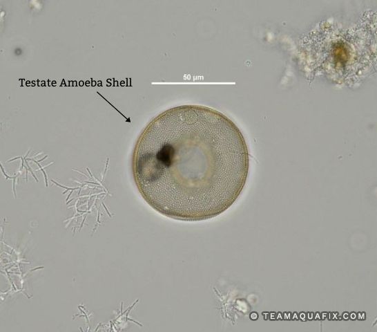
Testate Amoeba Aquafix from teamaquafix.com Paramecium under microscope 400x labeled. Education and information about the brain eating ameba naegleria fowleri that causes encephalitis and death including frequently asked questions, biology, sources of infection, diagnosis, treatment, prevention and control, and other publications and pertinent information for the public and medical professionals. This is a great view of cytoplasmic streaming, showing how an amoeba moves around by getting its cytoplasm to move to different parts of the cell, elongating. You may also want to learn more about actinophrys (small, heliozoan protists) here. Plantar warts are miniature growths appearing most commonly on the heel or other areas of feet or hands. Mikroskop 400x und ähnliche produkte aktuell günstig im preisvergleich. Arcella vulgaris (shown in the video above) is a testate amoeba. Hd amoeba at 40x 100x 200x and 400x youtube.
Amoeba using its pseodopodia to ooze forward (100x magnification, no sound).
Mikroskop 400x und ähnliche produkte aktuell günstig im preisvergleich. As such, they can only be viewed using a microscope. Amoeba (plural amoebas/amoebae) is a genus that belongs to kingdom protozoa. Amoeba using its pseodopodia to ooze forward (100x magnification, no sound). You may also want to learn more about actinophrys (small, heliozoan protists) here. Education and information about the brain eating ameba naegleria fowleri that causes encephalitis and death including frequently asked questions, biology, sources of infection, diagnosis, treatment, prevention and control, and other publications and pertinent information for the public and medical professionals. Video recorded by lee beavington at. Image of amoeba captured with the digital ba210 microscope at 100x magnification. Amoebas are a type of microscopic, unicellular protist and therefore will be best to see under a. Amoeba proteus is very well known for its extending pseudopodia. Believe it or not, this is a single cell! Plantar warts are miniature growths appearing most commonly on the heel or other areas of feet or hands. Amoeba under microscope 400x ad profundum.
Amoeba moves with their pseudopodia, which are a specialized form of the plasma membrane that results in a crawling motion of the amoeba under microscope. Biology protista amoeba malaria paramecium spirogyra.
